Radiographs are essential for the diagnostic and treatment planning process of orthodontics. Correct selection will help you deliver successful outcomes with aligner treatment. It is important to remember that radiographs for orthodontics are not ‘one size fits all’ – clinicians should not routinely take the same x-rays for every patient, selection must be patient-specific based on clinical findings and consider the risks and benefits of exposing the patient.
We all follow IRMER and understand for a radiograph to be justified we need to answer: – What is the objective of the exposure?- What is the benefit and detriment to the patient?- What alternative techniques could we use?
We must always ensure exposures are ALARP – all doses as low as reasonably practicable.
When it comes to orthodontic treatment, radiographs may be used at the initial examination, during treatment, at the end of active movement. They may also be helpful with interceptive treatment for example assessing eruption patterns or presence/position of canines if not palpable.
Types of radiographs we may take for orthodontic treatment include:
Intraoral Radiographs
Bitewings: Detailed images for caries and dental disease, detailed bone levels and helpful for checking restorations. Bitewings are a good supplement to panoramic radiographs, however they do not give information on the roots so not adequate alone for orthodontic treatment.
Periapicals: Helpful for checking root resorption and supplementing OPG findings. PAs can determine the presence and position of unerupted teeth and the presence of apical disease. Full-mouth PAs are rarely indicated as panoramic radiographs offer a similar amount of information with reduced exposure.
Upper Standard Occlusal Radiograph: This image shows the maxillary incisors and can be taken if there is potential for underlying disease or development anomaly in this area. Occlusal radiographs are helpful in assessing the position of Unerupted canines when used with a PA or and OPG using the parallax technique.
Extraoral Radiographs:
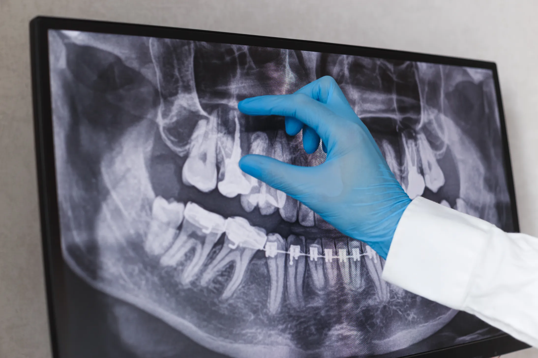
Dental Panoramic Radiographs (DPT): DPT’s can be taken to confirm the presence, position and morphology of unerupted teeth. One limitation of panoramic radiography is that the focal trough is relatively narrow, particularly in the incisor region, periapical radiographs may be needed to supplement this area. The FGDP in their 2013 selection criteria for dental radiography specified criteria for the use of panoramic radiographs which rules out the practice of taking panoramic radiographs for all new patients and screening asymptomatic patients, however they are appropriate when orthodontic treatment is being considered.
Lateral Cephalometric Radiographs: Cephalometric images may be used to aid diagnosis and treatment planning and can provide a baseline for monitoring progress. The patients who would benefit from lateral cephalometry are those with a skeletal discrepancy where the incisor relationship requires significant change, and it is unlikely that GDPs providing ‘cosmetic tooth alignment’ would benefit from taking these sorts of radiographs.
Sometimes a CBCT can be helpful for example assessing the position of unerupted teeth however generally panoramic and cephalometric radiographs appear to be sufficient in most orthodontic circumstances.
Once you have taken your selected radiographs, ensure you check all the images carefully and write a full report, including the justification, grade and report of findings for each radiograph.
A recommended reference is ‘Guidelines for the Use of Radiographs in Clinical Orthodontics’ – Isaacson et al, 2015. This document can assist you in the choice and timing of radiographs and is compliant with IRMER 2000.
Article by Lydia Sharples

